
Morphological ultrasound - what it is, when it is done, what is the price
A sonographic or ultrasound examination is one of the common examinations during pregnancy. Every woman is entitled to 3 ultrasounds, which are reimbursed by the health insurance company. The first is performed in the 12th, the second in the 20th and the third in the 30th week of pregnancy. Most mothers-to-be look forward to the ultrasound because it allows them to see the baby in the womb, preserve the memory of the period and at the same time find out if the fetus is developing properly.
 Author:
Jennifer Smith
Updated: 05.12.2023
- What is morphological ultrasound and when is it done?
- What does morphological ultrasound tell you about you and your baby?
- A morphological ultrasound will not tell you everything
- Is morphological ultrasound mandatory?
- 2D, 3D or 4D ultrasound
- Morphological ultrasound - experience
- The most frequent questions - FAQ
- Comments
Ultrasound examination is useful - it serves to check the course of pregnancy. The doctor checks not only the fetus, but also the environment in which it is located, especially the cervix. The first ultrasound examination reveals whether the baby is nestled in the uterus and confirms the number of fetuses (first-trimester screening), the second detects abnormalities in the structural development and growth of the child, as well as the position of the placenta (genetic ultrasound). In the 30th week, the function of the placenta and its placement, the amount of amniotic fluid and the size of the child are determined.
What is morphological ultrasound and when is it done?
It is a prenatal test that is performed in the 18th to 22nd week of pregnancy, the ultrasound is most often performed in the 20th week. It is used to check the size of the child and its body organs, possible birth defects are detected - scanning of fetal anomalies, but also the position of the placenta.
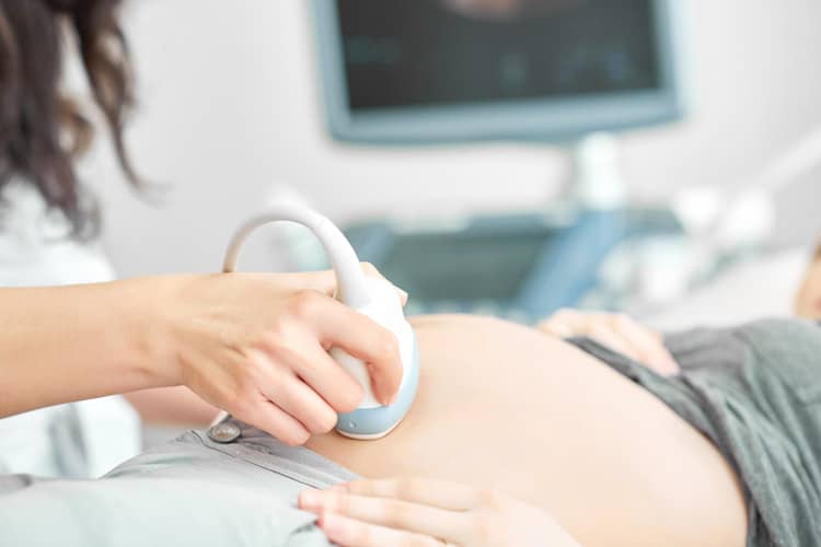
Pulses of sound waves pass from the transducer to the uterus, which bounce off the baby and create echoes. The sound waves are very quiet, they do not disturb the child. A computer turns these echoes into images that appear on the screen – bones are white, fluids are black, and tissue is mottled gray.

What does morphological ultrasound tell you about you and your baby?
Using morphological ultrasound, individual parts of the fetus and all organs are scanned. Under the microscope are organs such as the brain, heart, lungs, stomach, intestines, kidneys, liver, bladder, but also the development of the spine, limbs and parts of the baby's face. Morphological ultrasound also scans the placenta - its structure and location. Last but not least, this examination determines the amount of amniotic fluid. It can be used to determine the gender of the baby and a more precise date of birth. The examination takes approximately 30 minutes.
- Head: Head circumference (HC), biparietal diameter (BPD) and overall face are examined to see if the child has e.g. cleft lip.
- Spine: During the ultrasound, it is determined whether the spine is covered with skin, whether the vertebrae are forming and aligned.
- Heart: The size of the 4 chambers is examined, whether they are connected by valves and whether they open and close correctly with each heartbeat. The vessels connecting the heart are examined.
- Abdominal wall: It is determined whether it surrounds all internal organs, the circumference of the abdomen is measured.
- Stomach: The position of the stomach is monitored, it should be located just below the heart and should be filled with amniotic fluid.
- Kidneys and bladder: It is checked that they are properly formed and functioning.
- Limbs (arms, legs, hands, feet): It is checked that all the limbs are present, the femurs and humerus are measured so that the doctor can check that they are growing appropriately.
- Umbilical cord: The number of vessels in the umbilical cord is checked - there should be 2 arteries and 1 vein.
- Amniotic fluid: The measurement determines whether its quantity meets the standards.
- Placenta: The position of the placenta in the uterus and its distance from the cervix are recorded.
Advanced morphological ultrasound
An extended genetic ultrasound is recommended to expectant mothers if the prenatal screening showed bad values. If the gynecologist has any suspicions and, based on the results, recommends a more detailed ultrasound to the pregnant woman, the mother-to-be undergoes it free of charge (her insurance company pays for it). If you are interested in an extended morphological ultrasound, or if you want to perform an ultrasound screening beyond the scope of your health insurance, you must pay extra. The price varies depending on the selected action. It is about 50 to 100 euros.
With an extended morphological, genetic ultrasound, the profile of the face (mouth, lips, nose), individual parts of the brain, the state of important blood vessels and their flow are also monitored, the diaphragm and heart are examined in detail, and 3D/4D imaging is used. During the examination, anomalies are deliberately sought, which may indicate a serious developmental defect.
A morphological ultrasound will not tell you everything
Despite the fact that ultrasound can detect a number of birth defects (e.g. according to the number of fingers, the health of the heart, the state of the kidneys and bladder, or the state of the spine), it cannot detect all of them. At approximately the 20th week, the anomaly detection rate is 40 to 70%. You will usually find out the results of the examination directly at the clinic where the ultrasound is performed. If the screening reveals any anomalies, it is necessary to carry out additional examinations. Most often, it involves the collection of amniotic fluid or chorionic villi, which are used to determine whether the estimated diagnosis is correct.
Is morphological ultrasound mandatory?
A large ultrasound, fetal morphology or second-trimester screening is considered the most important ultrasound in pregnancy. The basic version is reimbursed by the health insurance company and every woman is entitled to three free ultrasounds during pregnancy (around 12, 20 and 30 weeks). If a woman does not want to undergo an ultrasound (e.g. she does not want to know in advance about possible disorders in the child's development), she can refuse the examination at her own risk.
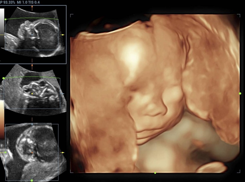
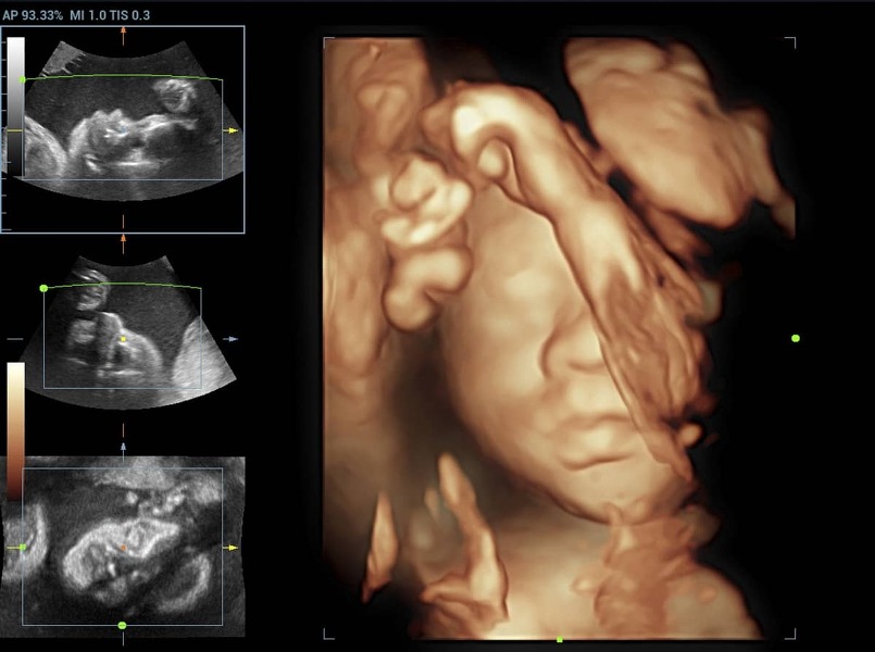
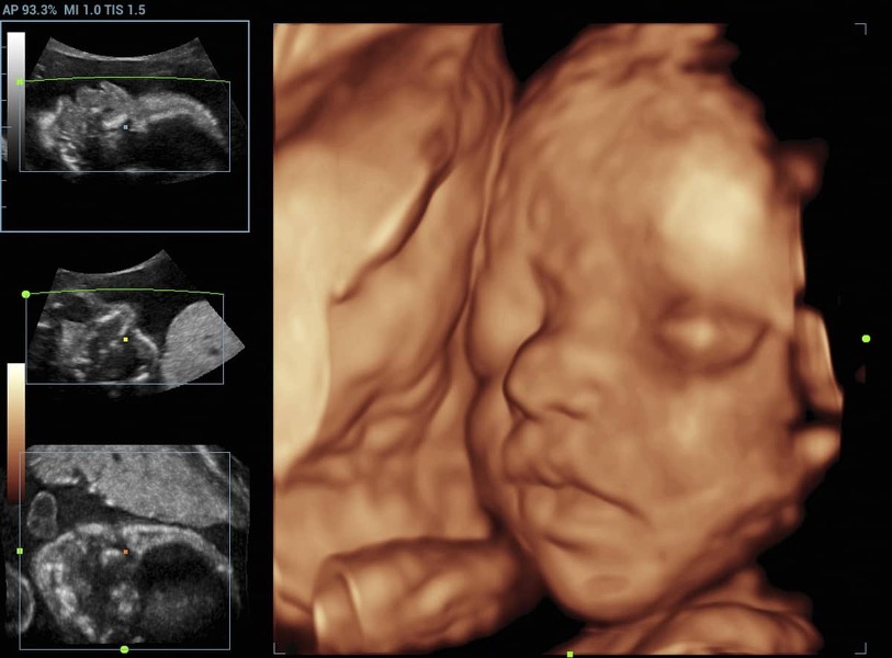
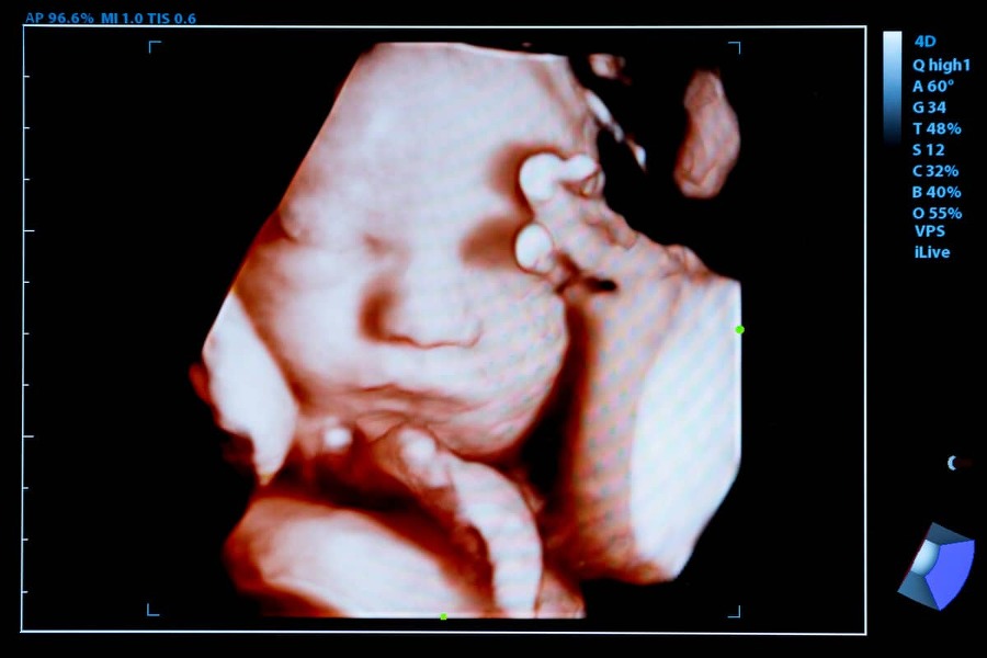
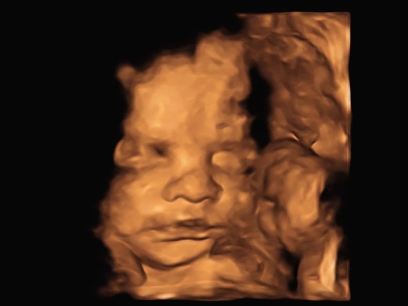
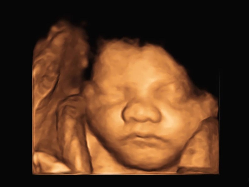
2D, 3D or 4D ultrasound
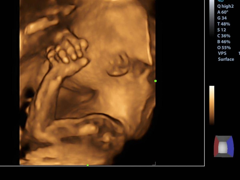
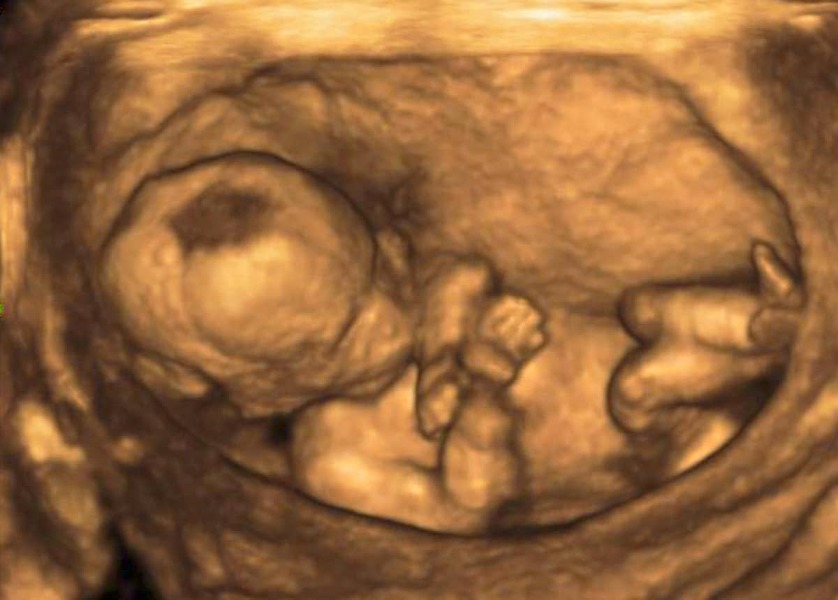
"Mandatory" prenatal ultrasounds are done in 2D form, it is enough to find out all the necessary data that the doctor needs to know about the fetus. It is the main diagnostic tool in ultrasound examination. 3D ultrasound represents a static image of the fetus, but in space. A three-dimensional image is achieved by computer processing a set of accumulated 2D images. Another possibility is a 4D display, which differs from a plastic 3D image by one additional factor – time/motion. You can see the image live, you can watch how the child moves and watch his facial expressions. 3D and 4D ultrasounds are not free.
Morphological ultrasound - experience
Moms shared their experiences with morphological ultrasound examination in discussions and forums. Most expectant mothers performed it in the 20th trimester. As they said, it is not a mandatory examination, but a woman is entitled to a large ultrasound. The doctor can use it to reveal the child's gender, fingers and toes, find out if the child's heart is working properly, his spine is developing properly, etc.
The most frequent questions - FAQ
Morphological ultrasound - yes or no? Expectant mothers are entitled to it, but they can also refuse it. If you are not interested in an ultrasound, ask your doctor about the procedure. The doctor can claim that you didn't go to the counseling office properly, which can affect the payment of the allowance when the child is born. We will be happy if you share your experience with a large ultrasound in the discussion and explain to other expectant mothers what its advantages and disadvantages are.
Where do morphological ultrasounds?
How to prepare for a morphological ultrasound?
Is ultrasound harmful during pregnancy?
Gallery
Pridať komentár